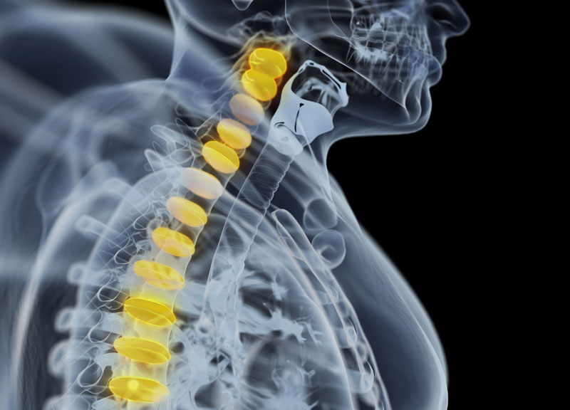
What is a CSF Leak?
Cerebral spinal fluid leak results when the fluid around the brain (called cerebral spinal fluid) leaks through a hole through the skull bone. This fluid can either drain from the ear or the nose, depending on where the skull bone is damaged.
Symptoms of CSF Leak
Drainage – Usually, patients complain of clear, watery drainage. This comes from only one side of the nose or ear. The drainage increases as the patient tilts the head forward or straining.
Other Symptoms – Headache, changes in vision, and loss of hearing
CSF leaks may be categorized into two groups:
1. Spontaneous Leaks – These may occur without any known cause.
2. Traumatic Leaks – The leaks are usually related to a history of surgery, head injury, tumors etc.
Diagnosis for CSF Leaks
History – The doctor will assess your medical history in detail.
Physical Exam – The doctor will perform a physical exam.
Endoscope – Usually the doctor will examine the nose using an endoscope. The physician may ask you to lean forward for several minutes. This will help him understand if leaning increases drainage. In case, the drainage can be collected, it will be sent for laboratory testing. There, the professionals will test the fluid to confirm that it is cerebral spinal fluid.
Imaging Studies – CT scans or MRIs may also be ordered to evaluate the patient for skull bone defects.
The Treatment Options for CSF Leak
The treatment for CSF Leak may be either medical or surgical. This depends on case to case.
Conservative Treatment
Doctors first suggest conservative treatment. It is prescribed for spontaneous CSF leak or head trauma.
Rest – The patient is required to follow bed rest for 1-2 weeks.
No Jerks – Patients are encouraged to avoid sneezing, coughing, and heavy lifting.
No Strain – Stool softeners are given to avoid straining.
Lumbar Drain – The doctors will place a lumbar drain in the lower back with an aim to decrease pressure of the CSF fluid around the leak area. This also helps the affected area to heal.
Surgical Treatment
This treatment option is considered when conservative treatment fails. The doctor may either use an external incision or endoscopic to gain access to the affected area. The graft material is placed in a fashion to close the skull base hole. Varied types of graft material available, such as tissue taken from other areas of the body. It may also be synthetic material. Nasal packing is often placed at the end of the case.
Schedule an Appointment Online
Submit an Online Request.
Call us now for an Appointment
To find a head and neck specialist for your requirements, get in touch with the Premier Brain & Spine.


